Cancer Screening
1. What is Cancer Screening?
2. Dangers of Screening: Overdiagnosis and Overtreatment
3. Breast screening: Overdiagnosis and Overtreatment
4. Prostate screening: Overdiagnosis and Overtreatment
5. Lung screening: Overdiagnosis and Overtreatment
6. Bowel Screening
7. Cervical Screening
8. The view of some health professionals
9. Some Solutions
10. Upcoming Technology?
1. What is Cancer Screening?
Checking for disease when there are no symptoms. Since screening may find diseases at an early stage, there may be a better chance of curing the disease. Examples of cancer screening tests are the mammogram (for breast cancer), colonoscopy (for colon cancer), and the Pap test and HPV tests (for cervical cancer).
2. Dangers of Screening: Overdiagnosis and Overtreatment
Definition of Overdiagnosis
Source: National Cancer Institute
Finding cases of cancer with a screening test (such as a mammogram or PSA test) that will never cause any symptoms. These cancers may just stop growing or go away on their own. Some of the harms caused by overdiagnosis are anxiety and having treatments that are not needed.
Some cancers that are diagnosed early do not develop symptoms requiring treatment, while others grow so slowly that the patient outlives the cancer and dies of other causes. Many of these are treated unnecessarily, leading to:
- Unnecessary tests and treatment
- Exposure to dangerous side-effects
- Radiation-induced cancers
- Mental and physical pain
- Infertility
- Inflated survival rates
This 2019 Opinion Article says: For cancer screening to be successful, it should primarily detect cancers with lethal potential or their precursors early, leading to therapy that reduces mortality and morbidity.
Screening programmes have been successful for colon and cervical cancers, where subsequent surgical removal of precursor lesions has resulted in a reduction in cancer incidence and mortality.
However, many types of cancer exhibit a range of heterogeneous behaviours and variable likelihoods of progression and death. Consequently, screening for some cancers may have minimal impact on mortality and may do more harm than good.
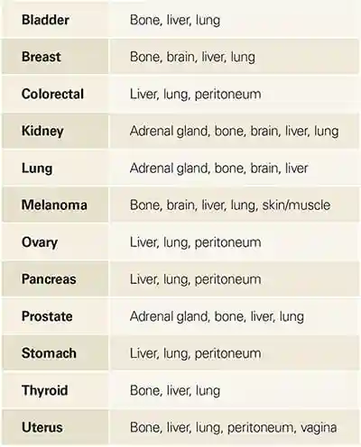
Prostate, Breast, Renal, Thyroid cancers, and Melanoma
This 2020 study found: After analysing changes in absolute lifetime risks for prostate, breast, renal, thyroid cancers and melanoma between 1982 and 2012, we estimated that 18% of all cancers diagnosed in women (ie, 11 000 diagnoses each year), and 24% of those in men (18 000 each year) are overdiagnosed cancers.
Lead time bias
A distortion overestimating the apparent time surviving with a disease caused by bringing forward the time of its diagnosis
The way statistics are manipulated is through the whole idea of early detection. Since cancer treatment success is measured on surviving five years after the first date of diagnosis, all we need to do in order to give the perception of greater success is to diagnose the patient earlier…
In his book The Cancer Code: A Revolutionary New Understanding of a Medical Mystery Dr Jason Fung explains:
Much of the benefit of screening is illusory due a phenomenon known as lead time bias. Imagine two women who both develop breast cancer at age sixty and who both die of the disease at age seventy. The first undergoes screening and finds the disease at age sixty-one. The other does not undergo screening and finds the disease at age sixty-five. The first woman “survived” cancer for nine years, whereas the second “survived” cancer for only five years. Screening improved cancer survival by an apparent four years, but this is only an illusion.

Figure: Lead time bias By Mcstrother – Own work, CC BY 3.0, https://commons.wikimedia.org/w/index.php?curid=15703636
A major study on cancer survival examined data over a 45 year period and found that “changes in 5-year survival rate over time bear little relationship to changes in cancer mortality.”
Some cancers spontaneously regress without intervention
This study looked at mammography screening in 229,256 Norwegian women (age 50-64 years).
Half of the women were invited for 3 rounds of screening over a six-year period, while the other half (the control group) were invited to undergo one screening at the end of a six-year period.
The study found that the cumulative incidence of invasive breast cancer was 22% higher in the screened group, leading the authors to conclude that some breast cancers detected by repeated mammographic screening would not persist to be detectable by a single mammogram at the end of 6 years. This raises the possibility that the natural course of some screen-detected invasive breast cancers is to spontaneously regress.
3. Breast screening: Overdiagnosis and Overtreatment
Swiss medical board call for end to mammography screening
In 2013, the Swiss Medical Board recommended that Mammography Screening be abolished in Switzerland ( The New England Journal of Medicine ) because of the emerging evidence that the harms of mammography outweigh the benefits.
The following is worth noting:
- Independent expert groups in Switzerland and France have recommended that breast screening be stopped or reduced substantially.
- A Canadian National Breast Screening Study found that Mammography did not reduce the number of deaths from breast cancer.
- A major 2017 study found that breast cancer screening leads to substantial overdiagnosis.
- There are safer breast screening methods available.
Some of the scientific evidence
A study published in 2017 in the New England Journal of Medicine involving thousands of breast cancer cases concluded that a significant proportion of tumors detected through mammography are not small because they are found early. Instead, the tumors are small because they are biologically prone to slow growth.
This study found mammograms can result in over diagnosis and unnecessary treatment.
This study involving 50-year-old Finnish women attending their first breast cancer screening found that mammograms can also be very painful. Sixty-one per cent reported painful and 59% uncomfortable mammograms (4% severely).
In this study 89,835 women, aged 40-59, were randomly assigned to mammography (five annual mammography screens) or control (no mammography). Conclusion: Annual mammography in women aged 40-59 does not reduce mortality from breast cancer…
This study conclused:
Screening with both MRI and mammography might rule out cancerous lesions better than mammography alone in women who are known or likely to have an inherited predisposition to breast cancer.
The authors of this study which looked at cancer overdiagnosis stated: We estimate the magnitude of overdiagnosis from randomized trials: about 25% of mammographically detected breast cancers…
This Article, published in 2012 in the New England Journal of Medicine, found: 31% of all breast cancers are over-diagnosed.
This 2016 review concluded:
· At least 20% of Breast Cancer patients, if left untreated would be alive after 5 years
· limited evidence suggests that around 10% of screen detected Breast Cancers may regress.
A Cochrane review of 10 trials involving more than 600,000 women showed no evidence that mammography screening reduced overall mortality (deaths).
This study says:
Invasive breast cancer
Of screening-detected invasive breast cancers, up to 30% might be ultra-low risk on the basis of their molecular profile. … Molecular classifiers are used to avoid chemotherapy, but could also classify tumours that have a very low or no risk of metastatic progression after simple excision, supporting the safe elimination of radiation therapy after lumpectomy in postmenopausal women, benefiting many women.
The National Cancer Institute’s Breast Cancer Screening (PDQ®)–Health Professional Version (updated June 1, 2018) includes the following :
Theoretically, in a given population, the detection of more breast cancers at an early stage would result in a subsequent reduction in the incidence of advanced-stage cancers. This has not occurred in any of the populations studied to date. Thus, the detection of more early stage cancers likely represents overdiagnosis. A population-based study in the Netherlands showed that about one-half of all screen-detected breast cancers, including DCIS, would represent overdiagnosis and is consistent with other studies, which showed substantial rates of overdiagnosis associated with screening.
This study sheds light on the need for new measures to avoid over diagnosis and unnecessary treatment.
The study authors say: A vast range of disorders—from indolent to fast-growing lesions—are labelled as cancer. Therefore, we believe that several changes should be made to the approach to cancer screening and care, such as use of new terminology for indolent and precancerous disorders. We propose the term indolent lesion of epithelial origin, or IDLE, for those lesions (currently labelled as cancers) and their precursors that are unlikely to cause harm if they are left untreated.
Ductal carcinoma in situ
Ductal carcinoma in situ is a pathological entity that is an unintended result of breast cancer screening, rarely diagnosed before screening was adopted. Diagnosis of ductal carcinoma in situ results in immediate treatment with aggressive locoregional therapy; however, the natural history of untreated ductal carcinoma in situ has never been elucidated. Results of studies suggest that only a subset of ductal carcinoma in situ progresses to clinically significant invasive cancer during a patient’s lifetime. The result of this historically aggressive treatment approach is that nearly 50 000 women per year in the USA are diagnosed and given treatment, 20 000 of whom undergo mastectomy, with a growing proportion opting for bilateral mastectomy.
[ Note: These women then attribute screening and treatment to saving their lives when in fact they did not need any treatment because the “cancer” detected was benign.]
New study finds breast cancer screening leads to substantial overdiagnosis
News Release issued by Nordic Cochrane Center
Women have been told for decades that they need to attend their routine breast screening appointments to detect early signs of breast cancer. However, a new major study, conducted by researchers at the Nordic Cochrane Centre in collaboration with researchers from Norway, looked into the effects of breast cancer screening programmes and have found a high risk of overdiagnosis and raise doubt about their benefits.
Risk of overdiagnosis outweighs the advantages of breast screening programmes
The cohort study, which was recently published in Annals of Internal Medicine, supports previous findings from the Cochrane review: Screening for breast cancer with mammography on the high risk of overtreating women and the doubts raised about the benefit of the intervention.
In an attempt to diagnose breast cancer early, a mammography screening very often detects small tumours that might not necessarily become malignant. In other words, the tumour would more than likely not be a life-threatening illness or health problem for the woman being treated. A breast cancer diagnosis is a life-changing event with profound implications for the psychological well-being and quality of life for women diagnosed and their families. They are subjected to invasive surgery, radiotherapy and sometimes chemotherapy, all of which are known to have serious, sometimes lethal, harms. The study also found that screening did not reduce the number of late stage tumours (those bigger than 2 cm), which means that breast screening is unlikely to reduce breast cancer mortality or lead to less invasive treatment.
Karsten Juhl Jørgensen, the lead author of the study explains, “In Denmark, we have the conditions to carry out a unique study as the screening programme was rolled out across the country in different regions at different times. We, therefore, had a control group of women of the same age during the same time period where the screening programme existed. This group of women were not screened over a 17-year period, making it possible for us not to have to speculate over what would have happened in the absence of screening”.
Additionally, breast screening leads to a 25 % to 50 % risk of being recalled due to a false positive test result if women attend a screening for the often recommended 20-year period. A false positive recall often means more mammograms and often biopsies. The time until a breast cancer diagnosis is excluded can be very stressful, and can have negative implications for many women’s quality of life even well beyond this period.
In his editorial on the study, Chief Medical Officer for the American Cancer Society, Dr. Otis Brawley recognises the need for accepting the limitations of mammography screening, “Acknowledging the existence of breast cancer overdiagnosis challenges the value of screening: it means that the benefits of breast screening have been overstated, and that some women who have been “cured” were harmed because they received unnecessary treatment”.
The main findings from the study are that:
- Screening did not reduce the number of late stage tumours (those bigger than 2 cm), which means that breast screening is unlikely to reduce breast cancer mortality or lead to less invasive treatment
- 1 in 3 breast cancers detected in women offered screening are likely overdiagnosed
Should mammography screening be stopped?
Independent expert groups in Switzerland and France have recommended that breast screening be stopped or reduced substantially. Elsewhere, guidelines are already changing as well, i.e. the American Cancer Society now recommend less frequent screening of a narrower age group than just two years ago. Our study supports this development.
The full report is titled: “Breast Cancer Screening in Denmark: A Cohort Study of Tumor Size and Overdiagnosis.” The authors are K.J. Jørgensen, P.C. Gøtzsche, M. Kalager, and P. Zahl.
For more information on breast cancer screening and the implication of the findings, please read the Cochrane Review on mammography screening or mammography screening information leaflet.
Karsten Juhl Jørgensen, the lead author of the study, can be contacted via email: kj@cochrane.dk.
This study says: We have also confirmed…our earlier finding that screening leads to more aggressive treatment, increasing the number of mastectomies by about 20% and the number of mastectomies and tumourectomies by about 30%…Furthermore, carcinoma in situ does not always develop into invasive cancer, but since these early lesions are often diffuse, women are sometimes treated by bilateral mastectomy. Therefore, the increase in surgery rates could also be an underestimate, since reoperations and operations in the contralateral breast seemed not to have been included.
This study of all women aged above 30 years (4.3 million, in 2000) in Norway and Sweden, says:… We conclude that after the introduction of screening programmes in Norway and Sweden one third of all cases of invasive breast cancer in the age group 50-69 are overdiagnosed—that is, without screening these cases would not have been detected during the patients’ lifetime.
Should you undergo regular breast screening using mammography?
That’s your call. The above information is for educational purposes only.
If you notice any symptoms or changes in your breasts, you should consult your GP without delay.
4. Prostate screening: Overdiagnosis and Overtreatment
In this study, Etzioni et al found that one in three prostate cancers diagnosed by screening for prostate specific antigen (PSA) is an over-diagnosis.
This study says:
With repeated prostate-specific antigen (PSA) testing and 10–12-core biopsy of the prostate, often done repeatedly, small, low-grade tumours are frequently detected. Attesting to the relatively low biological potential of these lesions are the 99% and 97% disease-specific survivals at 5 years and 10 years of follow-up, respectively, for men who are simply monitored and only given treatment if they have evidence of a grade or volume increase. Despite this indolent behaviour, greater than 90% of these tumours are treated with radiation or surgery, generating morbidities of treatment (eg, sexual, urinary, and gastrointestinal side-effects, in about 15–20% of patients), increased risk of secondary malignancies (with radiation), and increased cost.
5. Lung screening: Overdiagnosis and Overtreatment
This study says:
…70% of patients with stage I disease survive for 5 years, and 40% of those with stage II cancers. This finding has prompted major efforts to develop early detection programmes. However, lung cancer screening studies and autopsy reports suggest that a substantial subset of screening-detected cancers are indolent, even in the setting of a cancer type often thought to be aggressive, with 20–25% of cancer detected estimated to be overdiagnosed.
6. Bowel Screening
In Ireland – a BowelScreen Test Kit is offered free to men and women aged 60-69 by The National Bowel Screening Programme
This is a test you carry out in your own home.
You can also purchase the kit from PrivaPath Diagnostics
Other countries:
Test kits may be supplied by your Health Service or can be purchased in pharmacies and online.
7. Cervical Screening
What cervical screening is
Source: Health Services Executive
A cervical screening test checks the health of your cervix. The cervix is the opening to your womb from your vagina.
It’s not a test for cancer, it’s a test to help prevent cancer from developing.
Screening first looks to see if you have any of the high-risk types of human papillomavirus (HPV) that cause cervical cancer.
If HPV is found, your same test sample will be checked to see if you have any abnormal (pre-cancerous) cells in your cervix.
This is a new way of screening. It is called HPV cervical screening. It was introduced in Ireland in March 2020.
HPV cervical screening:
- is a better way of cervical screening
- prevents more cancers
- means some people will have fewer tests
If you have had a smear test in the past, having a cervical screening test will feel the same.
Key things to know about cervical screening
- It’s not a test for cancer, it’s a test to help prevent cancer from developing.
- All women or people with a cervix aged 25 to 65 should be invited for regular free screening by letter.
- During the screening test, a small sample of cells is taken from your cervix.
- The sample is tested for human papillomavirus (HPV).
- HPV can cause abnormal cell changes in the cervix.
- HPV is the main cause of cervical cancer.
- If your sample tests positive for HPV, we will check for abnormal cells.
- Abnormal cell changes are sometimes called pre-cancerous cells.
- In most cases, it takes 10 to 15 years for cells in the cervix to go from normal to pre-cancer to cancer.
- Finding HPV or abnormal cells early means you can be monitored or treated so they do not turn into cervical cancer.
- You’ll get your results by letter, usually about 4 to 6 weeks after your screening test.
What causes cervical cancer?
Source: The website of the National Cancer Institute
Nearly all cases of cervical cancer are caused by infection with oncogenic, or high-risk, types of human papillomavirus, or HPV. There are about 12 high-risk HPV types. Infections with these sexually transmitted viruses also cause most anal cancers; many vaginal, vulvar, and penile cancers; and some oropharyngeal cancers.
Although HPV infection is very common, most infections will be suppressed by the immune system within 1 to 2 years without causing cancer. These transient infections may cause temporary changes in cervical cells. If a cervical infection with a high-risk HPV type persists, the cellular changes can eventually develop into more severe precancerous lesions. If precancerous lesions are not treated, they can progress to cancer. It can take 10 to 20 years or more for a persistent infection with a high-risk HPV type to develop into cancer.
8. The view of some health professionals
In an interview with National Public Radio, Donald Lannin, a professor of surgery at the Yale School of Medicine, who led the study, said “It takes 15 or 20 years for [these small tumors] to cause any problems. And you can kind of imagine that a lot of patients will die of something else over that 15 or 20 years.
That means a significant proportion of women who get screened for breast cancer with a mammogram go through biopsies, surgery, chemotherapy, radiation — and experience a lot of stress — for tumors that may never pose a health threat.”
In the same interview, H. Gilbert Welch, a professor of medicine, community and family medicine at Dartmouth College, who was not involved in the study, said “It appears that screening disproportionately finds good cancers — cancers that may be better off not found,”.
“I think that we all need to realize that we’ve probably oversold the idea that looking for cancer early is the best way to avoid it,” Welch says. “Mammography’s a really close call. It’s a choice. We’ve exaggerated its benefit and we’ve sort of understated its harms.”
This is an eye-opening documentary on breast screening.
Get the full documentary at thepromisefilm.net
No Mortality Benefit of Breast Cancer Mammography Screening in 25-Year Follow-up of Canadian National Breast Screening Study
By Matthew Stenger
Source: The ASCO Post
Mammography Screening Reconsidered
Anthony B. Miller, MD
As reported in BMJ by Anthony B. Miller, MD, Professor Emeritus at the Dalla Lana School of Public Health, University of Toronto, and colleagues, the 25-year follow-up of the Canadian National Breast Screening Study has shown no mortality benefit of annual mammography screening for breast cancer compared with physical examination or usual care. Mammography screening was associated with substantial overdiagnosis…
…By the end of the 5-year screening period, there were 142 excess cases of breast cancer in the mammography group (666 vs 524). At 15 years after enrollment, the excess number of cancers in the mammography group became constant at 106 cancers. The excess number represents 22% (106/484) of all screen-detected cancers. Thus, there was one case of overdiagnosis of breast cancer for every 424 women screened by mammography during the trial.
The investigators concluded, “[O]ur data show that annual mammography does not result in a reduction in breast cancer specific mortality for women aged 40 to 59 beyond that of physical examination alone or usual care in the community. The data suggest that the value of mammography screening should be reassessed.”
Continue reading at The ASCO Post
9. Some Solutions
1. Reclassification of some “cancers”
Renaming low risk conditions labelled as cancer
Source: BMJ
Removing the cancer label in low risk conditions that are unlikely to cause harm if left untreated may help reduce overdiagnosis and overtreatment…Evidence is mounting that disease labels affect people’s psychological responses and their decisions about management options. The use of more medicalised labels can increase both concern about illness and desire for more invasive treatment.
This study says: A vast range of disorders—from indolent to fast-growing lesions—are labelled as cancer. Therefore, we believe that several changes should be made to the approach to cancer screening and care, such as use of new terminology for indolent and precancerous disorders. We propose the term indolent lesion of epithelial origin, or IDLE, for those lesions (currently labelled as cancers) and their precursors that are unlikely to cause harm if they are left untreated.
2. Alternative Screening Method: Digital Infrared Imaging (Thermography)
DII as a risk marker for breast cancer
Source: breastthermography.com
Studies show that an abnormal infrared image is the single most important marker of high risk for developing breast cancer, 10 times more significant than a family history of the disease (5). Consequently, in patients with a persistent abnormal thermogram, the examination results become a marker of higher future cancer risk (4,5). Depending upon certain factors, re-examinations are performed at appropriate intervals to monitor the breasts. This gives a woman time to take a pro-active approach by working with her doctor to improve her breast health. By maintaining close monitoring of her breast health with infrared imaging, self breast exams, clinical examinations, mammography, and other tests, a woman has a much better chance of detecting cancer at its earliest stage and preventing invasive tumor growth.
Angiogenesis, or new blood vessel formation, is necessary to sustain the growth of a tumor. Digital Infrared Imaging may be the first signal that such a possibility is developing .
See full article at breastthermography.com
Where can I get this test?
This test is now available at numerous medical facilities including:
Ireland
Hollywood Health (Northern Ireland)
United States
Breast Thermography (list of testing centers country-wide
See also
The National Cancer Screening Service (Ireland)
Good article on screening: HEALTHNEWSREVIEW.ORG
10. Upcoming Technology?
Molecular Breast Imaging: Potential New Tool for Detecting Cancers
Source: Oxford Journals
Mammography may be a “gold standard” for breast cancer screening, but for some women, its interpretation amounts to little more than a coin flip. In women with dense breasts, which contain more stromal and epithelial tissues than fat, interpretation of mammograms is difficult. Among these women, tumor detection rates with mammography barely exceed 60%. Both tumors and breast tissues appear white on a mammogram, but fat looks black. Clinicians can easily detect cancerous lesions against the darker fat backdrop, whereas dense tissues obscure tumors that could be life threatening.
Now researchers at the Mayo Clinic in Rochester, Minn., say that a different screening method— molecular breast imaging (MBI)— may offer a promising alternative for women with dense breasts, who make up much of the female population. Deborah Rhodes, M.D., an assistant professor at the Mayo Clinic, said roughly a quarter of all women older than 40 years have breast tissue that is more than 50% mammographically dense.
Compared with mammography, she said, MBI detected three times as many cancers among a population of 940 dense-breasted women who also had at least one additional breast cancer risk factor, such as a family history of the disease. These preliminary findings, announced in September at the American Society of Clinical Oncology’s 2008 Breast Cancer Symposium in Washington, D.C., came from an ongoing, comparative study of MBI and mammography that was launched at the Mayo Clinic in 2005. “We’re talking about a supplement to mammography that could be readily adopted by communities, instead of just major academic institutions,” said Rhodes, who is also the study’s principal investigator. “While [mammography is] terrific for some women, it doesn’t work well for others. For them, we need a better test, and that’s what MBI could provide.”
Read more at Oxford Journals
Deborah Rhodes: A test that finds 3x more breast tumors, and why it’s not available to you.
Page updated 2023